
Department of Nuclear Medicine in P. Hertsen Moscow Oncology Research Institute (MORI)
MORI’s Department of Nuclear Medicine performs a wide variety of nuclear medicine procedures.

MORI performs a high-tech diagnostics – positron emission tomography combined with computed tomography (PET/CT). PET/CT is a hybrid diagnostic technique of nuclear medicine, widely used in oncology practice all over the world.
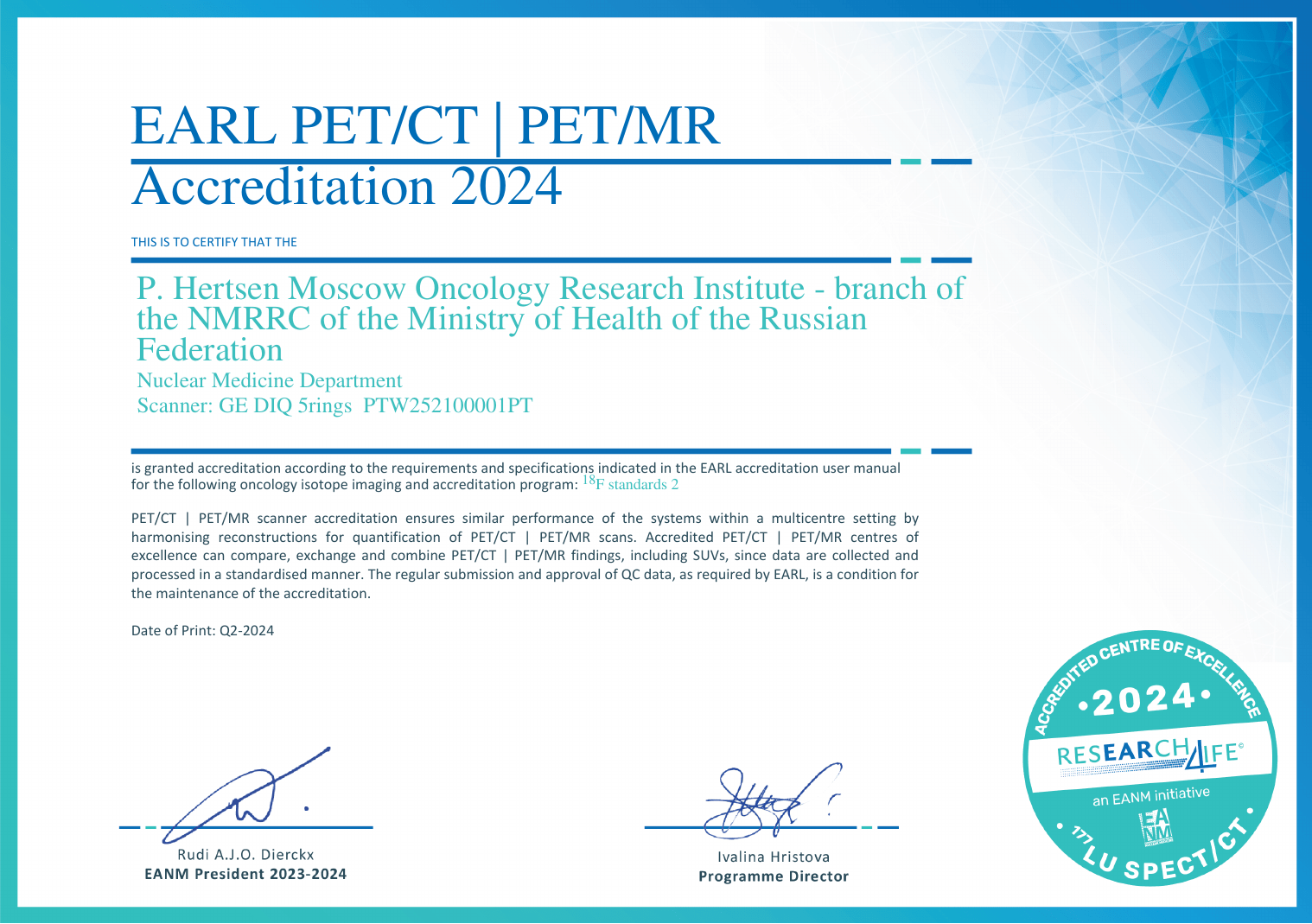
The National Medical Research Radiological Centre (NMRRC) of the Ministry of Health of the Russian Federation is the first center in the country accredited by the European Association of Nuclear Medicine (EANM) in the specialized EARL program, which aims to standardize the PET/CT procedure. This accreditation allows for reliable comparisons of studies performed on different scanners, which is especially important in assessing of treatment response in cancer patients.

Recommendations for patients undergoing [18F]FDG PET-CT
Подготовка к ПЭТ-КТ с [18F]ФДГ
Size: 655.62 KB
- Minimize Physical Activity: Limit physical activity and avoid sports
- Avoid Hypothermia: Take precautions to prevent hypothermia
- Dietary Adjustments: Replace foods high in carbohydrates (such as potatoes, beets, carrots, tomatoes, corn, cereals, baked goods, fruits, berries, juices, sauces, condiments, confectionery, and artificial sweeteners) with protein-rich options like meat, eggs, and green vegetables (including broccoli, asparagus, zucchini, and green beans), as well as cauliflower, mushrooms, cheese, and cottage cheese (without sugar and fruit additives)
- Avoid Certain Beverages: Do not consume alcoholic and sweet drinks, alcohol-containing medications, or milk. Non-carbonated water, coffee, and tea without sugar are allowed
- Avoid Steroid Medications: Refrain from taking steroid medications
- Fasting: Refrain from taking any food (including parenteral nutrition) for 6 hours before the scheduled time of the study
- Avoid Chewing Gum and Lozenges: Do not use chewing gum or lozenges on the day of the scan
- Dress Comfortably: Come to the department in comfortable clothing, if possible, without metal elements or jewelry
- Insulin Administration: Take the dose of insulin no later than 5 hours before [18F]FDG administration
- Oral Hypoglycemic Medications: Avoid taking oral hypoglycemic medications from the sulfonylurea and glinide groups 24 hours prior to [18F]FDG administration. It is advisable to stop taking biguanides as well; consult an endocrinologist for guidance on the temporary replacement of hypoglycemic medications
- Avoid Direct Contact: Avoid direct contact with the infant for at least 12 hours after the administration of [18F]FDG
- Timing of Breastfeeding: If possible, breastfeed or pump breast milk as close as possible to the time of [18F]FDG administration.
- Contrast Media Administration: The PET/CT scan is performed with intravenous administration of an iodine-containing contrast media if there are no contraindications.
It is mandatory for all patients to inform the medical staff about any allergic reactions in in their medical history, as well as any possible or confirmed pregnancy. Additionally, all patients undergoing PET/CT with intravenous administration of an iodine-containing contrast media must provide the results of serum creatinine analysis (no more than 1 month old).

Somatostatin Receptor Scintigraphy
This study allows for the visualization of tissues with a high expression of somatostatin receptors and is most widely used in the diagnosis of neuroendocrine tumors (NETs).
Indications for the study:
- Determination of the localization of the primary tumor and detection of metastatic lesions
- Prognosis of the effectiveness of therapy with somatostatin analogs
- Selection of patients for radionuclide therapy (PRRT)
- Evaluation of treatment efficacy
- Detection of recurrence and disease progression
The patient should provide the results of morphologic examination (including Ki-67) and laboratory diagnostics (creatinine, chromogranin A, calcitonin, serotonin, gastrin, etc.), information on any treatments received, as well as data of previous imaging (CT, MRI, ultrasound, scintigraphy, PET/CT). It is recommended to cancel somatostatin preparations (if tolerated) 2 days in advance for short-acting forms and 3-5 weeks in advance for long-acting forms, or to perform the study as close as possible to the date of the next injection. Additionally, it is preferable to follow a “liquid” diet for two days before the study and to use of laxatives the day before the study. The study is conducted in two stages: 2 and 4 hours after intravenous administration of radiopharmaceutical. Each stage lasts from 30 to 60 minutes.

Bone Scan
Bone Scan (or Bone scintigraphy) is more sensitive in detecting bone tumor lesions than traditional radiologic examination.
General indications for bone scan in cancer patients:
- Differential diagnosis of benign and malignant bone neoplasms
- Diagnosis of metastatic bone lesions
- Assessment of treatment efficacy for bone metastases
- Planning of radionuclide therapy with osteotropic radiopharmaceuticals
The patient should provide medical documentation on the referral diagnosis and data of previously performed studies: CT, MRI, PET/CT, SPECT/CT, scintigraphy, etc. on electronic media (CD/DVD or USB-drive) in DICOM format. Special preparation for bone scan is not required. The study is performed approximately 3 hours after intravenous administration of osteotropic radiopharmaceutical and takes about 30 minutes.

Bone SPECT/CT
If the results of bone scan are ambiguous, an additional hybrid technique – single photon emission computed tomography combined with computed tomography (SPECT/CT) – can be performed. This technique allows for reliable determination of the localization and clarification of the nature of the pathological process in the skeleton. This approach often eliminates the need for additional examination with other radiology techniques.
Bone SPECT/CT is usually performed on the same day as bone scan, does not require additional radiopharmaceutical administration, and takes 15 to 45 minutes.

Renal Scintigraphy
Renal scintigraphy is used to assess the functional state of the kidneys.
The patient should provide the results of biochemical blood analysis, including creatinine and urea levels, as well as previously performed kidney studies, such as ultrasound, radiography, MRI or CT (if available).
In preparation for the study it is necessary to follow a drinking regimen of 300-500 ml of non-carbonated water 30 min before the study. One day before the study it is necessary to stop taking diuretics. Renal scintigraphy is performed not earlier than 48 hours after intravenous administration of iodine-containing contrast media (CT, urography, etc.).
The study is performed immediately after intravenous administration of radiopharmaceutical and takes about 30 minutes.

Hepatobiliary Scintigraphy with SPECT/CT and Liver Functional Reserve Scoring
The study is used for preoperative planning of surgical treatment of liver tumor in order to predict the risk of liver failure.
It is necessary to avoid eating for 2 hours before the study. SPECT/CT is performed with intravenous administration of iodine-containing contrast media; therefore, the patient should provide the results of biochemical blood analysis, including the creatinine level (not more than 1 month old). The examination is performed immediately after intravenous administration of radiopharmaceutical and takes about 60 minutes.

SPECT Myocardial Perfusion Imaging
The study is performed to assess perfusion and contractility of the left ventricular myocardium.
The main indications for SPECT myocardial perfusion imaging:
- Diagnosis of ischemic heart disease
- Determination and assessment of the area of myocardial scar damage
- Assessment of antianginal therapy efficacy
The patient should provide the results of ECG, ECHO-CG.
The study involves a two-day protocol – one day at rest and one day with exercise stress test. On the day of the study it is necessary to avoid eating (for 2 hours before the study), smoking and caffeine-containing drinks.
Each stage of the study (at rest and with stress test) is conducted 40-50 minutes after intravenous administration of radiopharmaceutical and takes about 20 minutes.

SPECT/CT Parathyroid Imaging
The study allows to localize autonomously functioning parathyroid gland tissue (including ectopic tissue), which is extremely important for preoperative planning of minimally invasive surgical treatment of hyperparathyroidism.
The patient should provide the results of a neck ultrasound, as well as blood analysis results for ionized calcium and parathyroid hormone levels.
The study is conducted in two stages – 20 minutes and 120 minutes after intravenous administration of radiopharmaceutical. The duration of each stage is about 40 minutes.
MORI’s Department of Nuclear Medicine is equipped with:
- PET/CT-scanner GE Discovery IQ 5R
- Two SPECT/CT-scanners GE Discovery NM/CT 670 and Siemens Symbia Intevo Bold

About the Department of Nuclear Medicine
The Department of Nuclear Medicine performs studies on patients with cancer, as well as those endocrinologic, urologic and cardiologic diseases.
A wide range of nuclear medicine studies is performed daily, half of which are hybrid studies, such as SPECT/CT and PET/CT. Patients can receive expert consultations both in person and through telemedicine technologies.
Physicians of the department have extensive experience in the field of nuclear medicine and hybrid imaging. They actively participate in the scientific work of the NMRRC, are members of Russian and foreign scientific societies, and regularly make presentations on topical issues of nuclear medicine application in oncology. Thanks to the initiative of the department’s physicians, the topic of quantitative PET harmonization—requiring a multidisciplinary approach involving both nuclear medicine physicians and medical physicists—has begun to develop in the Russian Federation.
Khodzhibekova Malika Maratovna
Position: Nuclear Medicine Physician
Specialization: Radiology
Degree: doctor of medical sciences
Lazutina Tatyana Nikolaevna
Position: Nuclear Medicine Physician
Specialization: Oncology, Radiography, Radiology, Radiotherapy
Degree: candidate of medical sciences
Qualification: highest qualification category
Pylova Irina Valentinovna
Position: Nuclear Medicine Physician
Specialization: Radiography, Radiology
Degree: candidate of medical sciences
Qualification: highest qualification category
Leontyev Aleksey Viktorovich
Position: Chief of Department, Nuclear Medicine Physician
Specialization: Radiography, Radiology
Degree: candidate of medical sciences
Qualification: highest qualification category

Khalimon Aleksandr Igorevich
Position: Nuclear Medicine Physician
Specialization: Radiology
Khamadeeva Gulnara FaridovnaKhamadeeva Gulnara Faridovna
Position: Nuclear Medicine Physician
Specialization: Radiology
Khodakova Daria Yurievna
Position: Nuclear Medicine Physician
Specialization: Radiology
Nikiforuk Anastasia Igorevna
Position: radiologist
Specialization: Radiology