

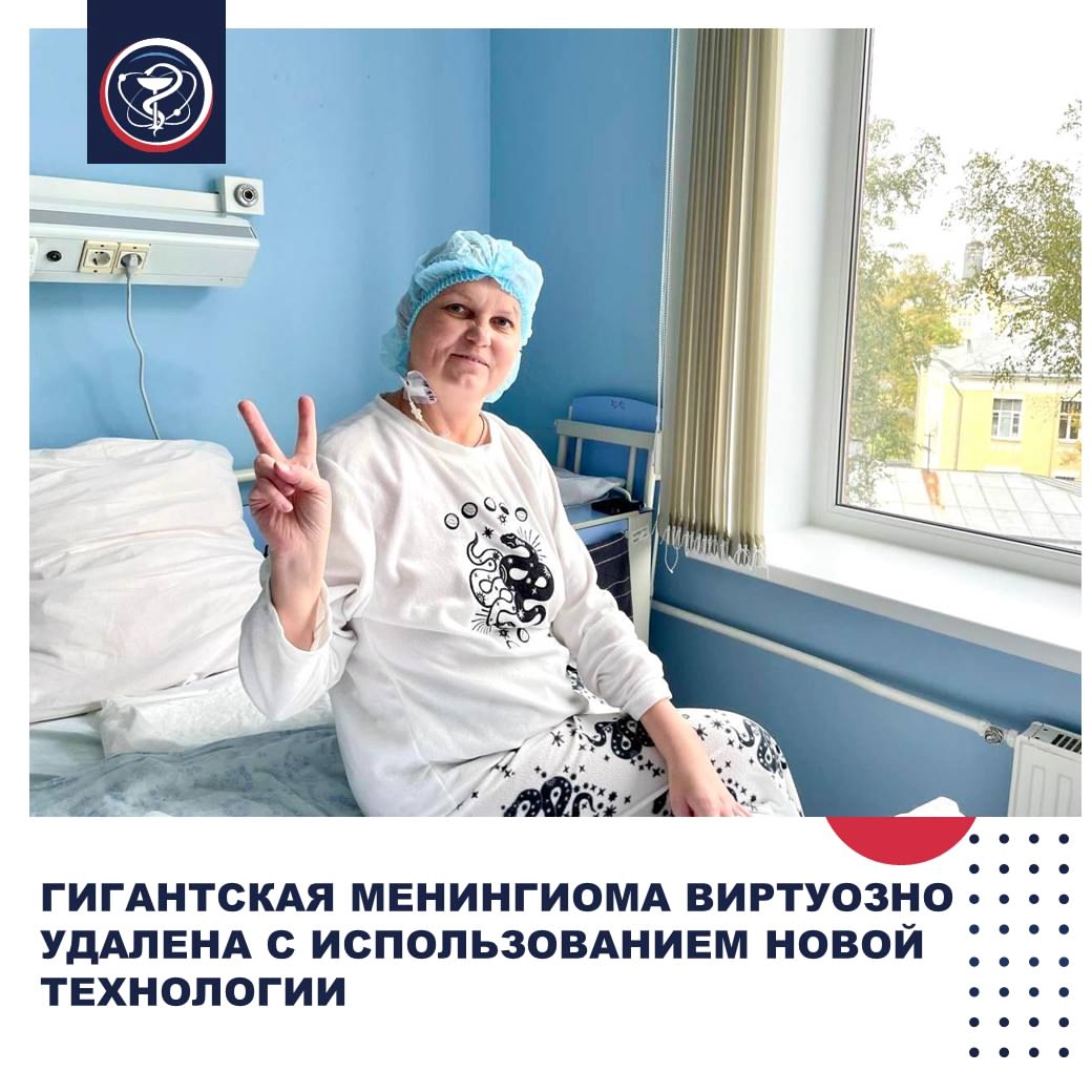

GIANT MENINGIOMA MASTERFULLY REMOVED USING NEW TECHNOLOGY
When Yulia came to the doctors at the National Medical Research Oncological Centre of the Ministry of Health of the Russian Federation, her tumor was already classified as giant.
“It measured over 6 cm and caused significant compression of the brain substance and displacement of the midline structures,” says the attending physician, senior research associate of the Neurosurgery Department, PhD Olga Kirsanova.
Although meningiomas are usually benign tumors, they can lead to paralysis, seizures, sensory and speech impairments. Moreover, they develop almost unnoticed.
“I felt my arm weakening,” recalls our patient from the Moscow region. “I thought I was just tired or it was something else, until one day I lost consciousness right on the street.”
Local doctors ordered an MRI and made a preliminary diagnosis. However, given the tumor size, they recommended consulting a specialized center. At the P. Hertsen Moscow Oncology Research Institute – the branch of the National Medical Research Oncological Centre of the Ministry of Health of the Russian Federation — the diagnosis was confirmed, and a two-stage tumor removal was proposed. The first stage aimed to reduce the risk of blood loss during the open brain surgery that followed.
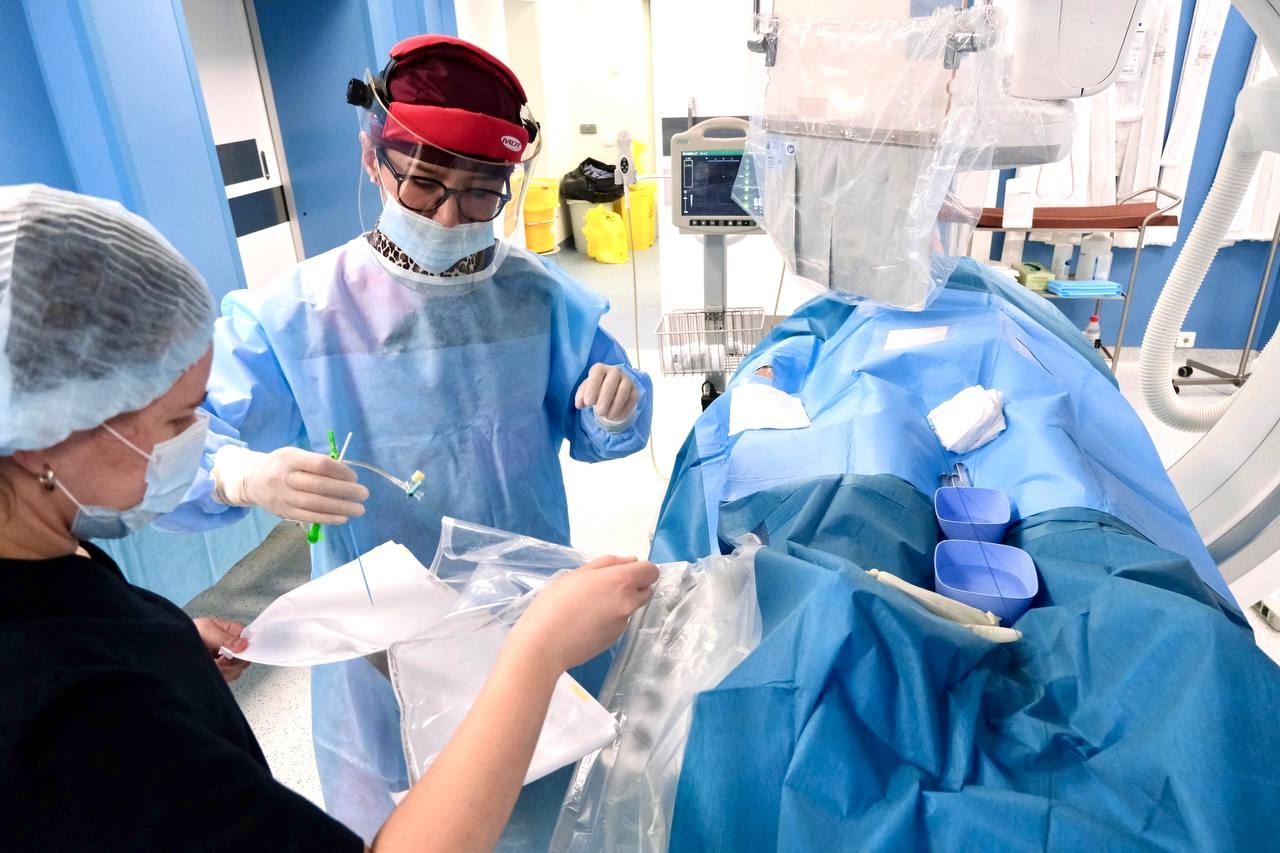
“The tumor had created an entire network of vessels supplying it for active growth. During open surgery, there is a real threat of heavy blood loss. Our task, using an endovascular procedure — blocking the vessels from within — was to close off these arteries,” explains head of the Department of Innovative Methods of X-ray Endovascular Surgery in Oncology at the P. Hertsen Moscow Oncology Research Institute, PhD Rimma Dugarova. “Here, we used an unusual technique: instead of standard embolic agents, we used a special adhesive compound. Its peculiarity is that it can block all small branches via the main vessel – in this case, the middle meningeal artery.”
Typically, with such a tumor volume, blood loss during surgery can reach 5 liters. In our case, it stopped at 200 ml. The use of adhesive composites is a known method in endovascular surgery but is rarely performed and requires special skills. Our specialists were assisted by Professor Kirill Orlov from the Federal Brain Center of FMBA, who visited the P. Hertsen MORI to share his invaluable experience.
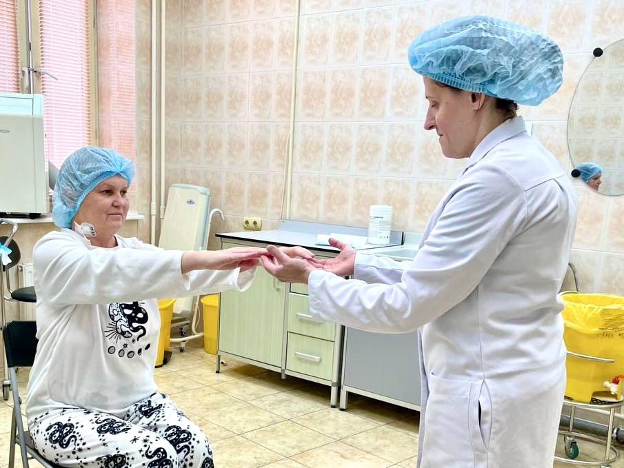
“During the tumor removal surgery, we successfully preserved the cerebral cortex and its blood supply, which allowed the patient to recover quite quickly with the help of our rehabilitation specialists.”
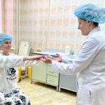
“Olga Nikolaevna is simply a magician; she gave me back my life,” Yulia says as she bids farewell. “God bless all the doctors of your Center!”