


THE STORY OF A LITTLE PATIENT: HOW THE NMRRC TEAM RESTORED HOPE
The New Year holidays have flown by. Today, all departments of the National Medical Research Radiological Centre are back to normal operations, with the same responsibility, attention and care for each patient. We are returning to our usual rhythm and will share with you, our readers, day by day, the most important, touching and remarkable stories from the life of the Centre – about people, doctors and the help that changes lives.
The Children’s Uro-andrology Department of the N. Lopatkin Scientific Research Institute of Urology and Interventional Radiology is renowned for its vast experience in treating the most complex urinary system diseases in children. One such case rightfully became part of the record of the most challenging surgical interventions of the past year.
A year ago, a three-year-old girl was admitted to the department with a rare congenital condition – bladder exstrophy. This is a condition in which the bladder develops outside the body, appearing «inside out» on the surface of the abdomen. Due to the condition, urine was constantly leaking, the skin was irritated, and the risk of kidney and urinary tract infections was increased. Additionally, children with this condition have a defect in the anterior abdominal wall and a separation of the pelvic bones, leaving the tissues under constant tension.
In the first days of life, at another clinic, the girl underwent her first surgery – an attempt was made to close the bladder and place it inside. However, the tissues could not withstand the tension and the bladder reopened. At about one year old, surgeons at another hospital performed a second surgery, but the tissues of the bladder and abdominal wall again separated. This is how the girl came to the N. Lopatkin Scientific Research Institute of Urology and Interventional Radiology. After a detailed analysis, pediatric urologists-andrologists decided to carry out treatment in two stages: in the first stage, the aponeurosis – the strong tissues of the anterior abdominal wall surrounding the bladder – was dissected to allow the bladder to stretch under abdominal tension, gradually grow and increase in volume.
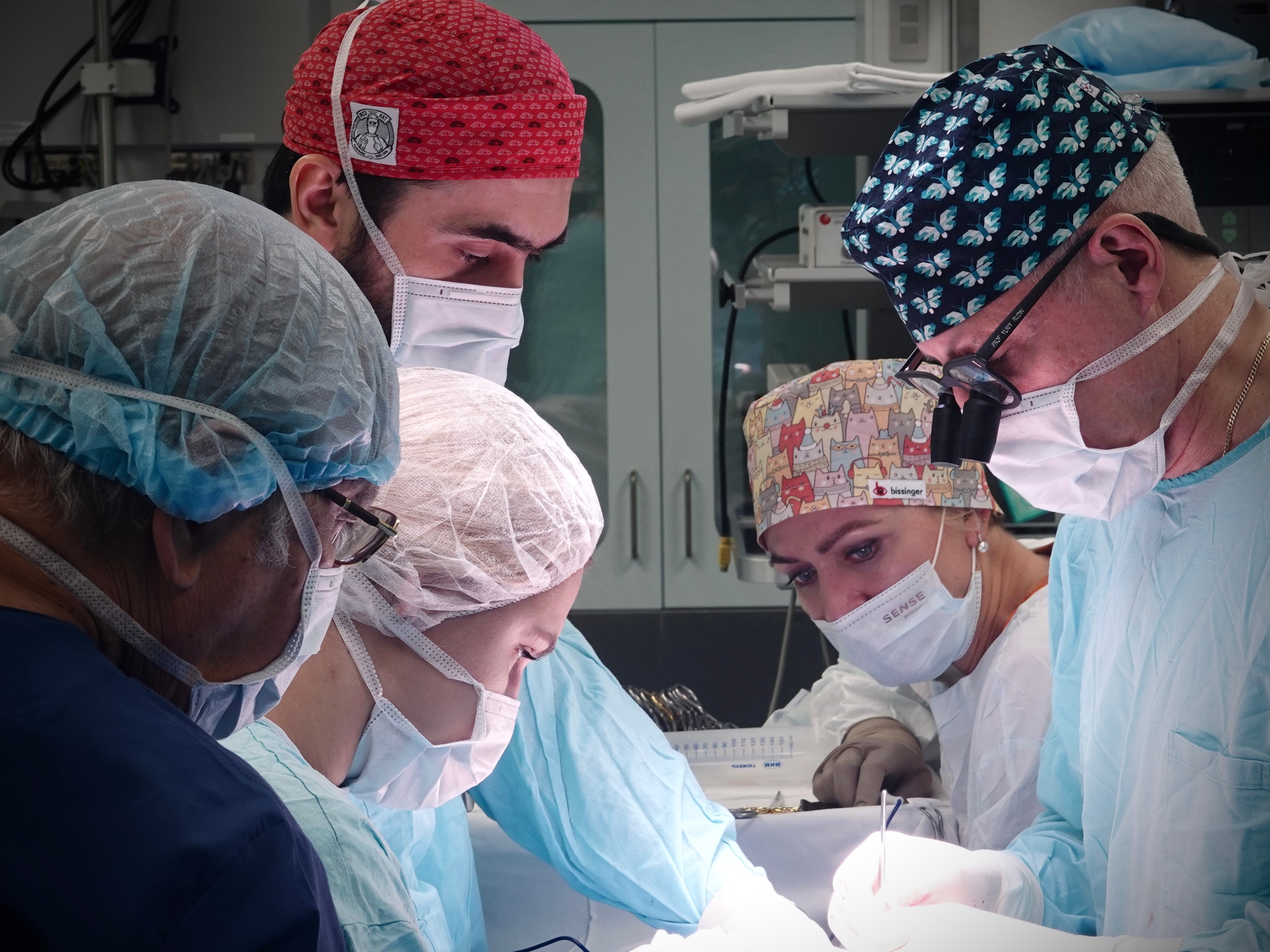
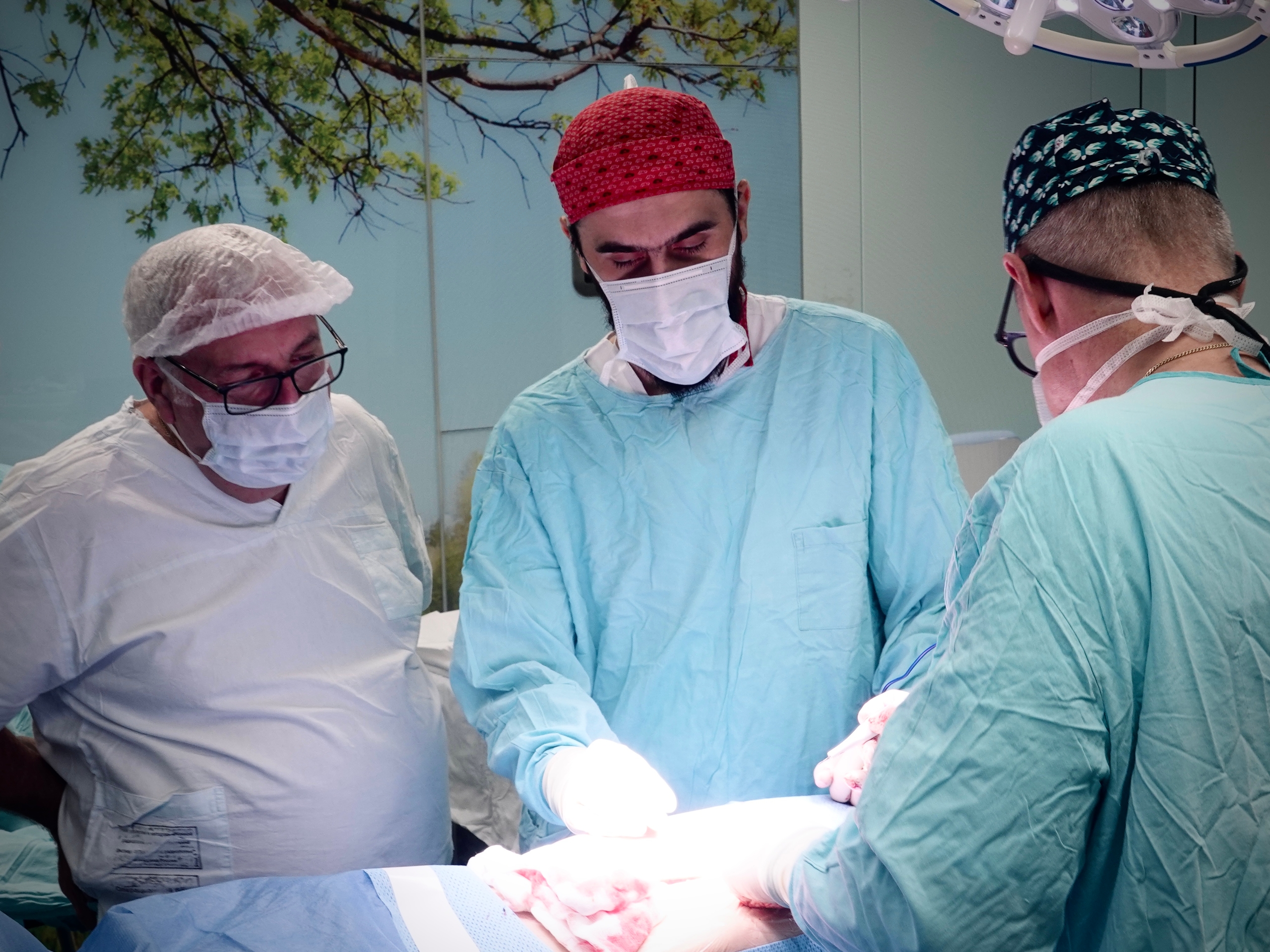
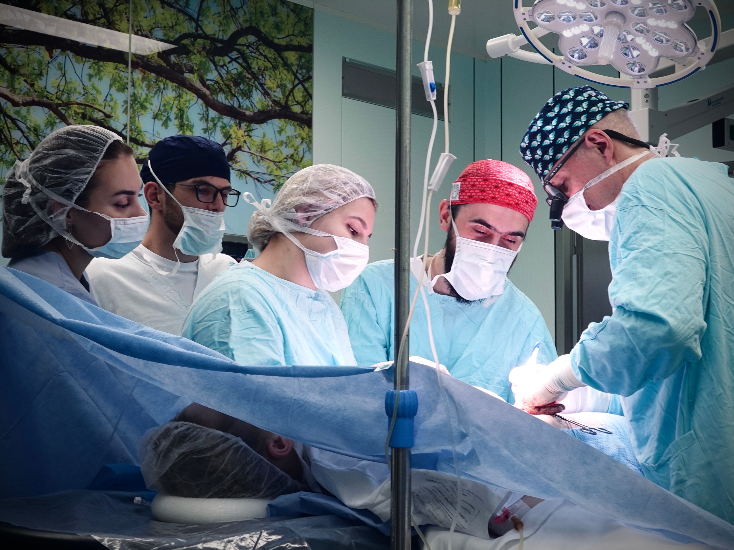
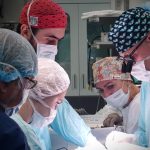
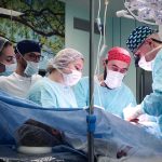
A year later, in 2025, when the bladder had grown larger, doctors performed the second stage of treatment: they closed the bladder again, transplanted the ureters to prevent urine from flowing back into the kidneys, and repaired its neck to improve urine retention. The key part of this stage was bilateral pelvic bone osteotomy: together with the pediatric trauma orthopedist, the surgeons brought together the pubic bones, minimizing their separation. «We achieved exactly the result we were hoping for», – explains the operating surgeon, head of the Pediatric Urology Team, Professor Yuri Rudin. «It was not a simple case, as numerous scars could have led to various complications, but we managed to help the child. If in the future additional corrective procedures are required to increase bladder volume or improve urine retention, we are always available».