

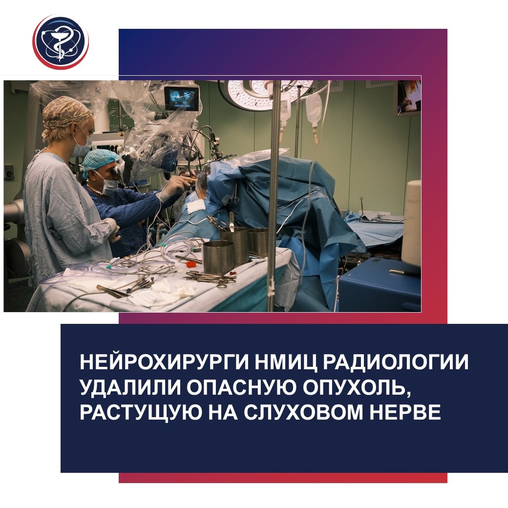
NEUROSURGEONS AT THE NATIONAL MEDICAL RESEARCH RADIOLOGICAL CENTRE REMOVED DANGEROUS TUMOR GROWING ON THE AUDITORY NERVE
For the past three years, Vadim had been complaining of hearing loss and was under the care of an ENT doctor. However, there were no improvements. He was referred for an MRI of the brain, which revealed a vestibular schwannoma on the vestibulocochlear nerve. This nerve is responsible for transmitting auditory impulses in our body. Although vestibular schwannoma is a benign tumor, its location in the cerebellopontine angle can cause significant issues, including severe complications leading to disability and potentially the patient’s death.
Small tumors (up to 2.5 cm in diameter) are successfully treated with radiosurgery by oncologist-radiologists using Gamma Knife and Cyber Knife systems. For larger tumors, surgery is indicated. Removing such a tumor is classified as a level 5, the highest category of surgical complexity, and may involve severe complications, including fatal ones. However, if surgeons can completely remove the tumor, it generally does not recur, and the patient recovers.
The challenge in surgery is the tumor’s location: it is situated in a difficult-to-access and anatomically complex area at the base of the skull, known as the cerebellopontine angle. “This area houses critical neurovascular structures of the brainstem, where the centers controlling breathing and cardiovascular activities are located. Additionally, very important cranial nerves, such as the hypoglossal, facial, auditory, and trigeminal nerves, as well as arteries supplying these vital centers, are also located here,” explains A.M. Zaitsev, PhD, head of the Neurosurgery Department at P. Hertsen Moscow Oncology Research Institute – branch of the Federal State Budgetary Institution of the “National Medical Research Radiological Centre” of the Ministry of Health of the Russian Federation. “Intraoperative injury to any of these structures can result in severe disability for the patient.”
Thanks to the implementation of the national healthcare project, the operating rooms at the Center are equipped with modern specialized equipment, allowing surgeons to perform necessary intraoperative neurophysiological monitoring of vital functions and assess the integrity of cranial nerves during surgery.
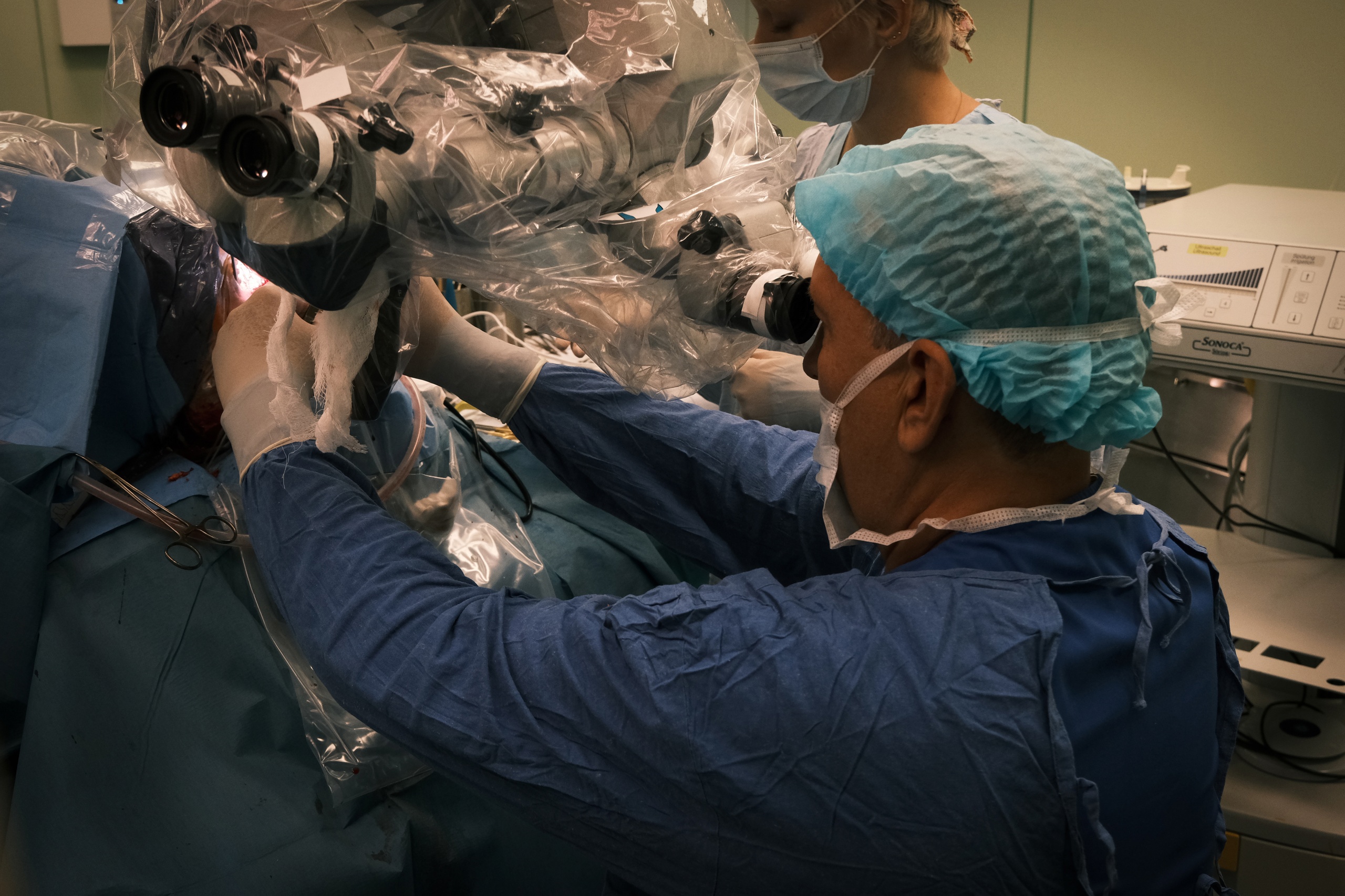
“A neurophysiologist helps us determine the location of these critical neurovascular structures in relation to the tumor and remove it without damaging these crucial structures,” explains Anton Mikhailovich. “He identifies the evoked potentials (signals) from the areas where the surgeons are working and indicates how close or far the surgeon is from these structures, guiding us on where we can operate confidently and where extra caution is needed.” The surgeons’ goal is to remove the tumor as radically as possible while preserving all the aforementioned neurovascular structures and, consequently, the patient’s quality of life. It’s important to note that all surgical manipulations are performed under a microscope, and the nerve’s thickness usually does not exceed 1 mm.
The surgery lasted about 4 hours and was complication-free. After a follow-up MRI and the results of a planned morphological study, the doctors will determine the next steps in the patient’s treatment: whether radiosurgery is needed or if dynamic monitoring will suffice. The neurosurgical team successfully completed the planned volume of the surgical treatment, and now they hope that Vadim’s life is no longer in danger.