


Innovative Method for Early Immunological Diagnosis of Metastases
The A. Tsyb Medical Radiological Research Centre (MRRC) – the branch of the FSBI “National Medical Research Radiological Centre” (NMRRC) of the Ministry of Health of the Russian Federation has unique laboratory studies that allow the detection of circulating tumor cells, the identification of which is related to disease prognosis and may trigger metastasis formation. According to international research data, it is possible to predict the occurrence of clinically significant metastasis using methods of so-called «liquid biopsy», which detect tumor markers (individual circulating tumor cells, circulating tumor DNA) in peripheral blood.

A. Tsyb MRRC Contact Center
This is important to know for the patients with a cancer diagnosis!
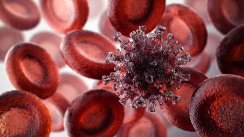
What is a circulating tumor cell (CTC)?
A tumor is a dynamic biological formation in which numerous processes are constantly occurring: some tumor cells die during its growth, some actively proliferate, and some viable cells seem to slough off from the surface of the tumor and undergo certain changes that biologists and oncologists call epithelial-mesenchymal transition. As a result of such changes, a tumor cell can penetrate through the wall of a blood vessel directly into the bloodstream, and such a cell is called a circulating tumor cell (or, abbreviated, CTC). It is precisely CTC that are currently considered one of the parameters of liquid biopsy, the detection of which in the peripheral blood of an oncology patient may be important for determining further treatment strategies.
It is important to know!
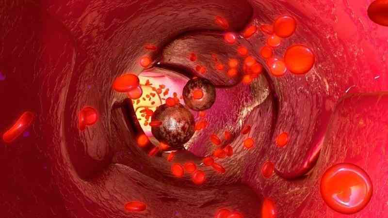
Not every circulating tumor cell (CTC) can lead to metastasis, and most of them will be inactivated by cells and biologically active components of the blood. Nevertheless, some CTCs (usually individual hyperactive cells) travel through the blood and lymphatic vessels to the brain, lungs, liver, bone tissue, spine, and kidneys. Once they reach internal organs and settle there, the cells acquire the status of disseminated tumor cells (DTCs), where they can remain in a dormant state for a long time. Biologists characterize the dormant state of such cells based on an assessment of the cell cycle phase (the period when the cell is not actively dividing). However, upon a certain stimulus or signal, these cells awaken, begin to divide, and form clinically significant metastases detectable by standard examination methods.
It is believed that cells that have undergone epithelial-mesenchymal transition possess a number of special properties — and, most importantly, they may be resistant to chemo-radiotherapy.
Considering the biological heterogeneity of CTCs (some of them are viable, some are not, some CTCs may transition into DTCs, some will be removed from peripheral blood, and some will acquire aggressive properties), a comprehensive assessment of CTCs over time is important, that is, during and after the completed comprehensive treatment. Upon detection of circulating tumor cell, the patient can be conditionally classified into a high risk group for metastasis. Modern laboratory methods, in particular the method implemented at the A.Tsyb MRRC, based on the immunological detection and characterization of circulating tumor cells and disseminated tumor cells, allow for the determination of both the quantity of CTCs and the assessment of their quality and viability, that is, factors associated with the likelihood of developing distant clinically significant metastases of the disease.

In which diseases can CTCs be detected?
Circulating tumor cells can be detected in most epithelial tumors (adenocarcinomas and squamous cell carcinomas), as well as in melanoma. Currently, their greatest prognostic significance has been established for breast cancer, prostate cancer, and colorectal cancer. Threshold values are considered to be <5 CTCs per 7.5 ml of blood for breast and prostate cancer, and <3 CTCs for colorectal cancer. It has been established that an increase in the number of CTCs above these values is associated with disease prognosis and shortens the relapse-free survival period.
When can DOK be evaluated?
Assessment of minimal bone marrow involvement (that is, evaluating the amount of DTCs) is important for patients suffering from skin/mucosal melanoma or breast cancer, as metastatic bone marrow involvement is not uncommon in these conditions. Detecting such subtle involvement at an early stage of the disease may be crucial for preventing extensive tumor spread.
How to submit material for CTC and DTC research?
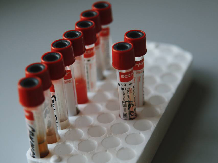
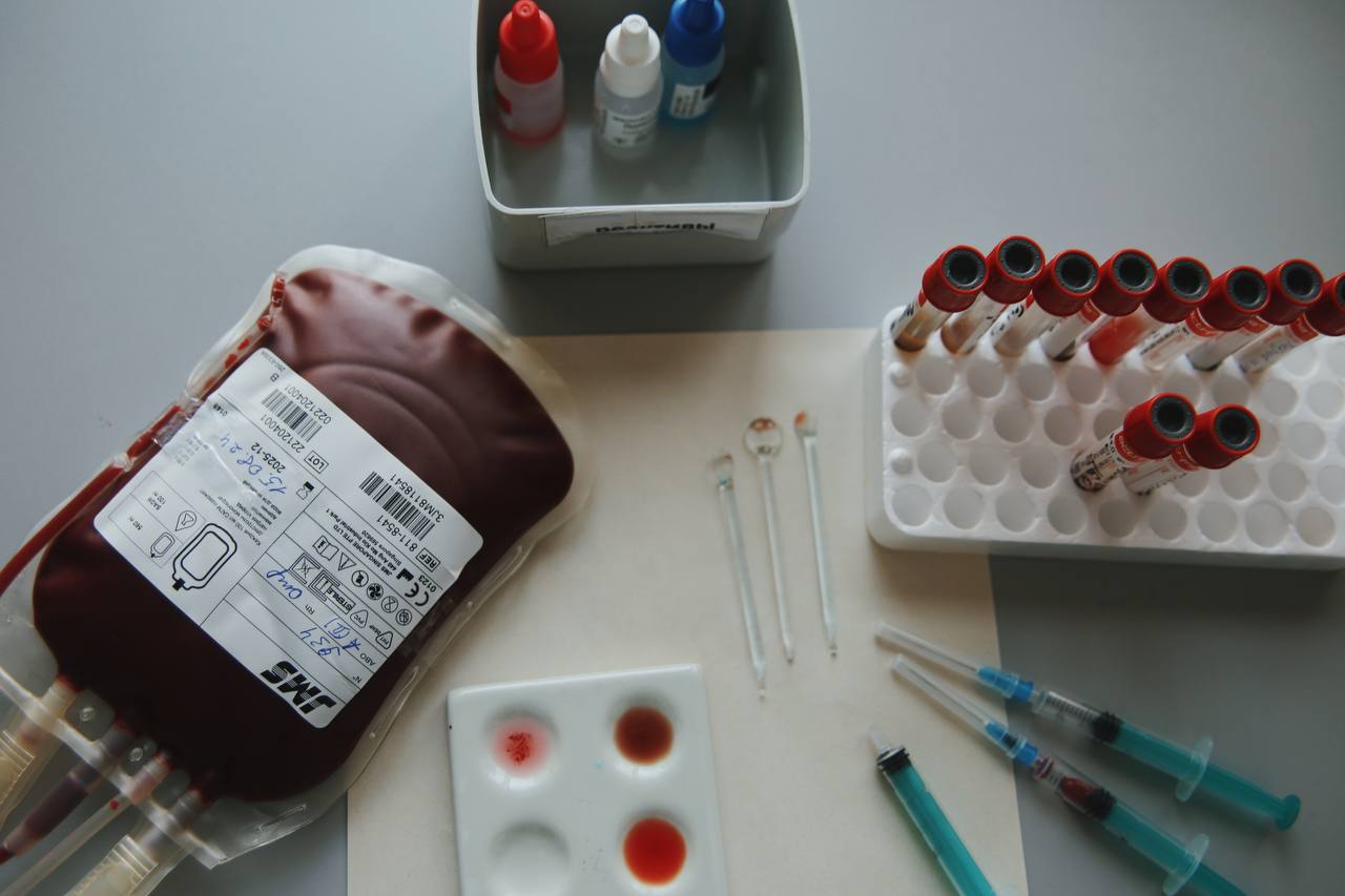
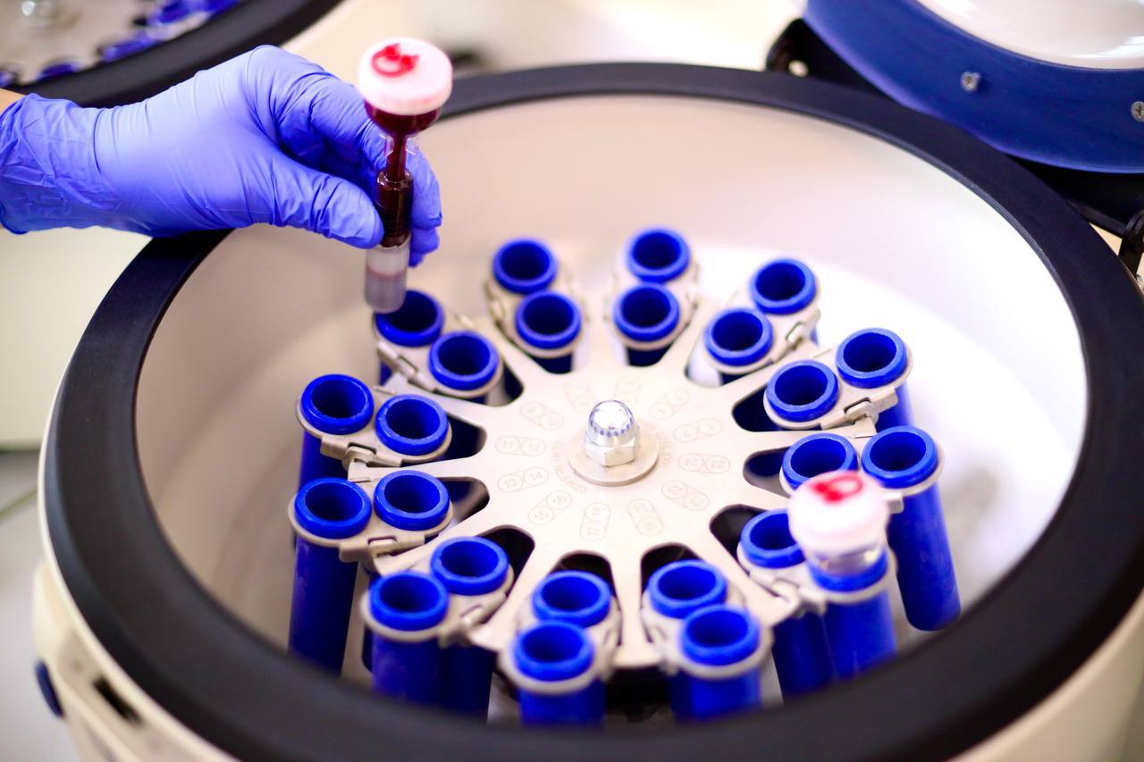
Modern laboratory diagnostic methods allow for the detection of both individual tumor cells and their clusters using immunological techniques. For CTC assessment, the patient provides a blood sample from a peripheral vein into a special tube containing a preservative (K3EDTA) that prevents blood clotting. The blood will be processed in a special way, live cells (leukocytes) will be isolated, and among them, cells with a specific set of protein markers on their surface will be identified (a complex of epithelial markers is evaluated, and hematopoietic cells, i.e., blood cells, are excluded from the analysis). These protein markers are detected by incubating the sample cells with reagents labeled with special fluorescent tags, which, if present, will be detected by the instrument using laser detection (multiparametric flow cytometry method).
In order to identify these individual cells (their frequency is low and sometimes does not exceed 1 cell per 100,000 blood cells), it is necessary to examine at least 10 ml of peripheral blood. The study of CTCs is quite labor-intensive, as it requires analyzing a large number of blood cells to detect true individual CTCs; therefore, it takes at least 3–4 days.
The methodology for assessing the number of DTCs is similar to the methods for detecting CTCs; however, in the case of DTCs, the analysis is conducted not on peripheral blood but on a bone marrow aspirate. Bone marrow is usually highly cellular, and to detect CTCs, 0.5 ml of liquid bone marrow obtained through an aspirational biopsy is sufficient. This procedure is performed by a hematologist and can be done on an outpatient basis, meaning during a clinic visit.
For CTC analysis, there is no need to observe a fasting condition.
What the definition of CTC provides
Through blood analysis and liquid biopsy methods, it is possible to:
- predict the likelihood of metastasis development — both in the near and distant future;
- monitor the effectiveness of comprehensive treatment (that is, to most fully assess the extent of the disease or the effectiveness of the ongoing treatment).

Exact name of the study CTC and DTC
Immunological study of bone marrow cells (disseminated tumor cells), 6-color flow cytometry;
Immunological study of tumor cells in peripheral blood (circulating tumor cells), 6-color flow cytometry.
Recommendations for Patients
Tests for the detection of CTC and DTC are conducted at the Department of Laboratory Medicine in A. Tsyb MRRC.
CTC detection tests are conducted from Monday to Thursday, no appointment is needed. DTC tests are conducted by appointment only, which can be scheduled by phone: +7 (800) 250-87-00.
You will receive your results within 4–5 working days!