
PET/CT
Positron emission tomography combined with computed tomography (PET/CT) is a hybrid nuclear medicine technique widely used in oncology worldwide. It allows for both structural and molecular imaging in a single study. This comprehensive approach to imaging enables more effective diagnostics in oncological disorders.

P. Hertsen Moscow Oncology Research Institute (MORI) contact center:
N. Lopatkin Scientific Research Institute of Urology and Interventional Radiology (SRIUIR) contact center:

- High Accuracy
- Early Detection
- Available for patients from Russia and foreign patients
PET/CT is a high-tech imaging modality that complements conventional imaging (such as CT, MRI, ultrasound, etc.) and, in some cases, can fully replace them. It enables the assessment of tumor prevalence, detection of recurrence, planning of treatment and assessment of treatment response. Patients can undergo PET/CT at two branches of the NMRRC.

Principle of PET/CT
During the procedure, the patient receives a small amount of a radiopharmaceutical, the distribution of which, depending on its composition, reflects certain processes occurring in the body. Visualization of these processes is made possible by the radioactive isotope that is a part of the radiopharmaceutical, which emits gamma particles that are captured by PET-scanner. CT allows for the localization of molecular changes in the patient’s organs and tissues detected by PET.
[18F]fluorodeoxyglucose, or [18F]FDG, is the main radiopharmaceutical used for PET/CT imaging worldwide, particularly in oncology. Its accumulation in organs and tissues correlates with the level of glucose metabolism, which is typically elevated in malignant tumor cells of most types.

Common Clinical Indications for [18F]FDG PET/CT
- Differentiation of benign from malignant lesions
- Identification of the primary tumor site in cases of metastatic disease as the first manifestation of cancer or when the patient presents with a paraneoplastic syndrome (CUP)
- Tumor staging
- Assessment of treatment response
- Evaluation of the nature of post-treatment residual changes
- Detection of tumor recurrence, including cases with elevated tumor markers
- Selection of the area in the tumor lesion that is most informative for biopsy
- Planning of radiotherapy
Contraindications for [18F]FDG PET/CT
While there are no absolute contraindications for [18F]FDG PET/CT, the following relative contraindications should be considered:
- Pregnancy (suspected or confirmed)
- Acute medical conditions
- Severe somatic disorders
- Infection or inflammation in acute phase
- Hyperglycemia (blood glucose level above 11 mmol/L)
- Conditions that may hinder the correct performance of the study (e.g., neuropsychiatric disorders, including claustrophobia, acute pain, etc.)
[18F]FDG PET/CT should be performed not earlier then:
- 7 days after biopsy
- 21 day after chemotherapy (if the interval between administrations is shorter, schedule as close to the next one as possible)
- 2 months after surgery
- 3 months after radiotherapy

Recommendations for patients undergoing [18F]FDG PET-CT
Скачать рекомендации по подготовке к процедуре ПЭТ/КТ с 18F – ФДГ
Size: 336.85 KB
Recommendations for 24 Hours before study:
- Minimize Physical Activity: Limit physical activity and avoid sports
- Avoid Hypothermia: Take precautions to prevent hypothermia
- Dietary Adjustments: Replace foods high in carbohydrates (such as potatoes, beets, carrots, tomatoes, corn, cereals, bakery, fruits, berries, juices, sauces, condiments, confectionery, and artificial sweeteners) with protein-rich options like meat, eggs, and green vegetables (including broccoli, asparagus, zucchini, and green beans), as well as cauliflower, mushrooms, cheese, and cottage cheese (without sugar and fruit additives)
- Avoid Certain Beverages: Do not consume alcoholic and sweet drinks, alcohol-containing medications, or milk. Non-carbonated water, coffee, and tea without sugar are allowed
- Avoid Steroid Medications: Refrain from taking steroid medications
Recommendations for the Day of study:
- Fasting: Refrain from taking any food (including parenteral nutrition) for 6 hours before the scheduled time of the study
- Avoid Chewing Gum and Lozenges: Do not use chewing gum or lozenges on the day of the scan
- Dress Comfortably: Come to the department in comfortable clothing, if possible, without metal elements or jewelry
Recommendations for Patients with Diabetes Mellitus undergoing [18F]FDG PET-CT
- Insulin Administration: Take the dose of insulin no later than 5 hours before [18F]FDG administration
- Oral Hypoglycemic Medications: Avoid taking oral hypoglycemic medications from the sulfonylurea and glinide groups 24 hours prior to [18F]FDG administration. It is advisable to stop taking biguanides as well; consult an endocrinologist for guidance on the temporary replacement of hypoglycemic medications
Recommendations for Breastfeeding Patients undergoing [18F]FDG PET-CT
- Avoid Direct Contact: Avoid direct contact with the infant for at least 12 hours after the administration of [18F]FDG
- Timing of Breastfeeding: If possible, breastfeed or pump breast milk as close as possible to the time of [18F]FDG administration
- Contrast Media Administration: The PET/CT scan is performed with intravenous administration of an iodine-containing contrast media if there are no contraindications
It is mandatory for all patients to inform the medical staff about any allergic reactions in their medical history, as well as any possible or confirmed pregnancy. Additionally, all patients undergoing PET/CT with intravenous administration of an iodine-containing contrast media must provide the results of serum creatinine analysis (no more than 1 month old).

How is [18F]FDG PET/CT performed?
The patient should arrive at the reception of the Department of Nuclear Medicine at the scheduled time. After filling out the necessary documentation, the patient will be accompanied to the procedure room, where the technologist will perform the required measurements, place a peripheral venous catheter, and administer [18F]FDG.
After [18F]FDG administration, the patient should remain in a comfortable semi-reclining position in a special relaxation chair for approximately one hour. This time interval is required for radiopharmaceutical distribution and obtaining correct PET/CT results. During this period, it is forbidden to talk, listen to music, read, or use mobile devices.
Immediately before the PET/CT scan, the technologist will ask the patient to void the bladder and will position the patient on the scanner table.
The PET/CT scan lasts between 15 to 45 minutes, during which the patient should remain still and follow all instructions from the technologist.
If there are no contraindications, the patient will receive an iodine-containing contrast media at the end of the PET/CT scan.
After the study, the patient may leave the department only if authorized by the medical staff.
During the next 24 hours after [18F]FDG administration, it is necessary to:
- Limit close contact with others, especially small children and pregnant women
- Increase fluid intake to 2 liters
- If possible, avoid salty foods, as this can lead to [18F]FDG retention in the body
If needed, a certificate confirming the administration of the radiopharmaceutical can be issued upon request on the day of the PET/CT scan.
The medical report will be provided no earlier than 48 hours after the PET/CT.

Documents Required for [18F]FDG PET/CT
The patient must provide the following documents:
- Passport or other ID and its scan copy
- Medical documentation regarding the referral diagnosis
- Data from previously conducted imaging, including reports (if available), such as CT, MRI, ultrasound, PET/CT, SPECT/CT, and scintigraphy, in electronic format (CD/DVD or USB drive) in DICOM format

[18F]PSMA-1007 PET/CT for Patients with Prostate Cancer
The main indications for [18F]PSMA-1007 PET/CT in prostate cancer are:
- Primary staging (if the results may affect treatment) in patients with unfavorable intermediate or high risk (Gleason score 7 (4+3) or higher, more than 50% positive cores on biopsy, and/or T2c or higher, and/or PSA level ≥20 ng/mL), as well as in cases of equivocal results from conventional imaging at any risk level
- Biochemical recurrence (elevated PSA level: ≥0.2 ng/mL after radical prostatectomy confirmed by serial repeated measurements; elevated PSA level: ≥2 ng/mL or more above nadir after radical radiotherapy) and biochemical persistence (PSA level above 0.1 ng/mL for more than 6 weeks after radical prostatectomy)
- Biochemical signs of castration-resistant prostate cancer (presence of three consecutive PSA level elevations one week apart, with an increase of 50% above the nadir in two measurements, with PSA level >2.0 ng/mL) and no signs of progression by conventional imaging (preliminary bone scan, CT, and MRI are obligatory).
- Planning and assessment of treatment response of PSMA-targeted radioligand therapy
In any other clinical situation it is necessary to consult a nuclear medicine physician from the MORI’s Department of Nuclear Medicine by phone: +7 (495) 945-87-18
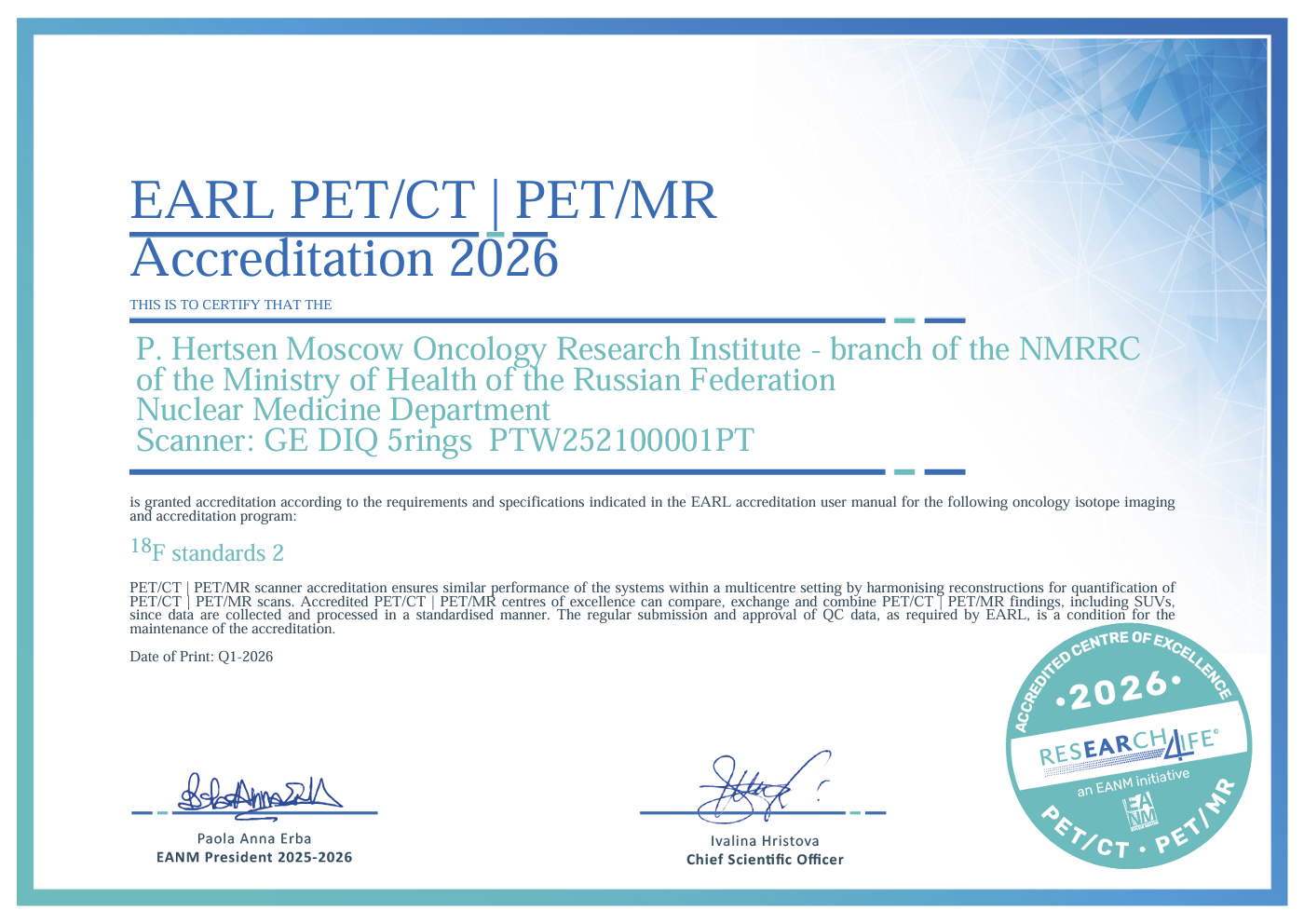
EARL Accreditation by the European Association of Nuclear Medicine (EANM)
In 2010, the European Association of Nuclear Medicine (EANM) launched a specialized EARL program aimed at quantitative PET harmonization. This initiative allows for reliable comparisons of studies performed on different scanners, which is especially important when assessing treatment response in cancer patients, as diagnostic results significantly impact further treatment strategies and the prognosis of the disease.
In 2023, the GE Discovery IQ 5r PET/CT scanner installed in the MORI’s Department of Nuclear Medicine became the first EARL-accredited scanner in the Russian Federation. In 2024, the second EARL-accredited scanner in the country was also the GE Discovery IQ 5r, installed in the Department of Nuclear Medicine at the N. Lopatkin Scientific Research Institute of Urology and Interventional Radiology (SRIUIR), another branch of the National Medical Research Radiological Centre of the Ministry of Health of the Russian Federation.
In 2024, EARL launched a new program aimed at promoting high quality and harmonizing approaches in theranostics. The program includes three levels of certification, and as of early 2025, it encompasses only the first, introductory level, which involves gathering relevant information from interested centers to establish best practices in theranostics. In January 2025, MORI became the first accredited theranostics center in the Russian Federation to join the new EARL program.