

What does an MRI of the pelvis show and what is included?
Magnetic resonance imaging (MRI) of the female pelvis is prescribed for the primary diagnosis of diseases of the reproductive system (body and cervix, ovaries, fallopian tubes, vagina, vulva), bladder and intestinal tract, assessment of anatomical features, and is also performed to assess the effectiveness of treatment.

N. Lopatkin Scientific Research Institute of Urology and Interventional Radiology Contact Center:
+7 (499) 110 40 67
MRI of the pelvis for diagnostics of gynecological diseases
- shows changes in the structure of organs and tissues at the scanning level;
- helps to distinguish benign from malignant tumors;
- identifies pathological processes at an early stage;
- helps in preoperative planning;
- evaluates the effectiveness of the treatment;
- allows to avoid invasive interventions (for example, diagnostic laparoscopy).
Another important advantage of MRI of the pelvis is multi-plane visualization: images can be obtained in any section (longitudinal, transverse, frontal), which gives the doctor a complete picture of the anatomy of the pelvic organs and their possible disorders.
Magnetic resonance imaging of the pelvis provides detailed images of internal organs and tissues, revealing even minor changes. Among its advantages are high accuracy, safety (no ionizing radiation) and excellent visualization of soft structures.
This is an indispensable method in modern gynecology, oncology, urology and surgery, helping to quickly and safely make an accurate diagnosis and choose effective treatment.
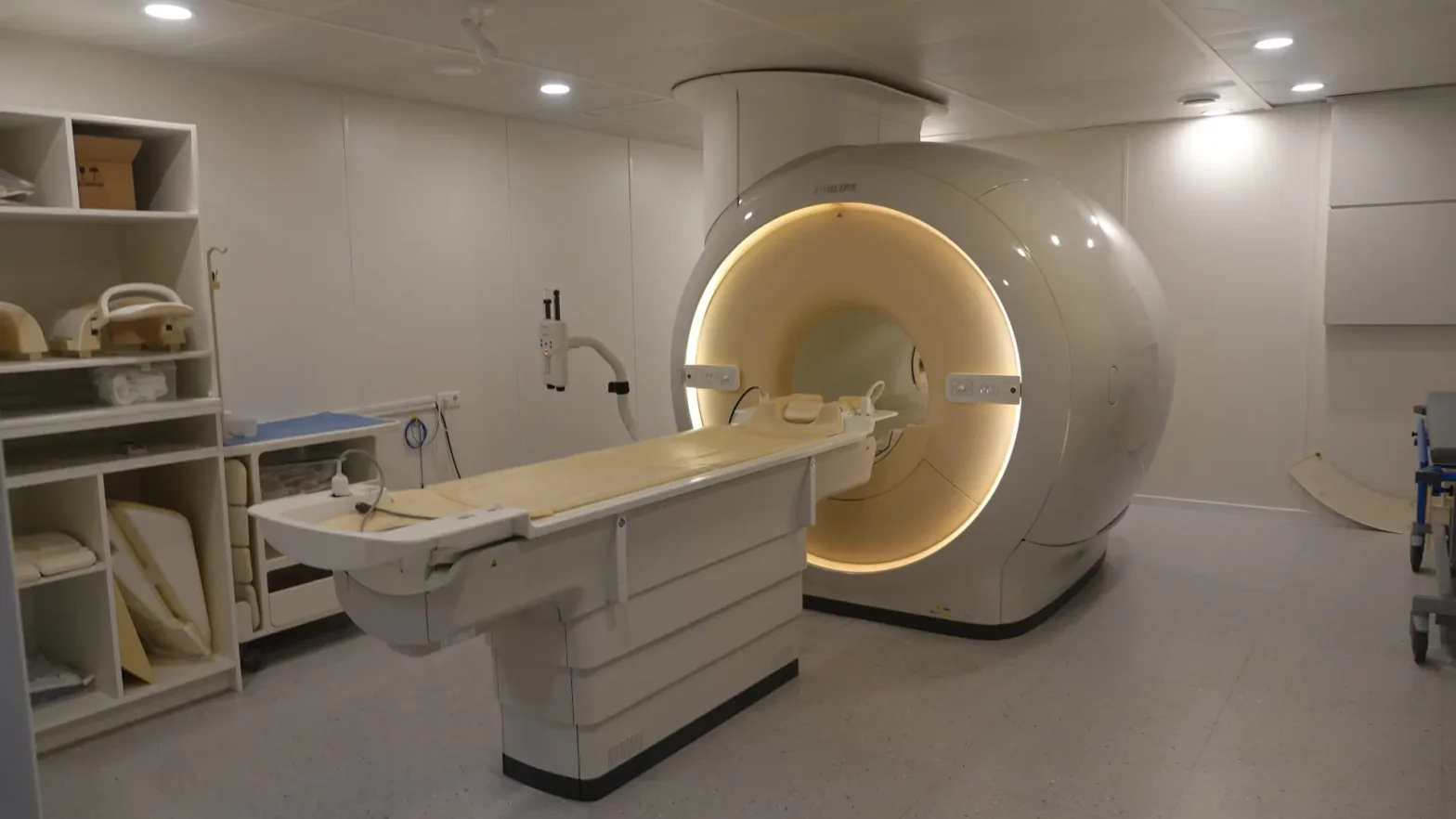
How is a pelvic MRI performed?
A pelvic MRI procedure is performed in a special diagnostic device, an MRI scanner, which is a large cylindrical unit with a movable table. This equipment creates a powerful magnetic field, with the help of which detailed images of internal organs are formed.
The question of how to prepare for the examination often arises, and here it is important to take into account small nuances.
First, the patient will be asked to fill out an informed consent, which specifies contraindications for the examination (for example, the presence of a pacemaker, hearing implant, etc.), remove metal objects and change into a pre-prepared disposable shirt.
Before the start of the pelvic MRI, the patient is explained what will happen and how long it will take.
The patient lies down on a soft movable couch, puts on headphones (to eliminate noise during the operation of the tomograph). Medical staff helps to correctly position the body and fixes the position to avoid movement during the scan. If necessary, you can use rollers or straps for additional comfort and stability.
The couch is smoothly moved into the tunnel of the tomograph. Rhythmic sounds and clicks are heard during the operation of the device – this is absolutely normal. The patient may be offered headphones or earplugs to reduce noise. Important: it is necessary to remain still throughout the procedure, as movement can worsen the quality of the images and distort the results.
In some cases — if there is a suspicion of neoplasms, metastases or inflammatory processes — the doctor may recommend MRI of the female pelvis organs with contrast. Particular attention is paid to how to prepare for MRI of the pelvis in the presence of contrast enhancement. The contrast agent (gadolinium-based) is administered intravenously and helps to more clearly visualize the vessels and pathological foci. The drug is well tolerated and is excreted naturally through the kidneys.
After the scan is completed, the couch returns to its original position, and the patient can stand up calmly. If there is no contrast, no recovery or observation is required – you can immediately return to your normal life. If contrast is used, it is recommended to drink more fluids during the day to speed up the elimination of the drug.
On average, 30–40 minutes.
With contrast — up to 60 minutes.
MRI of the pelvis is painless, calm, and fairly quick for the patient, the main thing is to follow the doctor’s recommendations.
Proper preparation for MRI of the pelvis will help to obtain more accurate and high-quality images.
What conditions can a pelvic MRI show?
A pelvic MRI is prescribed in cases where the most accurate and detailed information about the state of the reproductive system and adjacent organs is required. This method is especially useful when other diagnostic methods do not provide a complete picture of the patient’s condition.
- Discomfort
MRI of the pelvic organs in women is performed in the presence of pain or discomfort in the lower abdomen, especially when other examination methods do not give a clear result. This highly accurate method allows for a detailed study of the internal organs and surrounding tissues, identifying inflammatory processes, neoplasms, cysts, adhesions and other hidden pathologies. - Menstrual irregularities
Late, excessively heavy or painful menstruation, as well as bloody discharge between cycles – all this may indicate abnormalities in the structure of the uterus, endometrium or ovaries. MRI of the pelvis allows you to determine the causes of hormonal imbalance or identify anatomical abnormalities. - Suspected gynecological diseases
MRI of the pelvic organs (PMO) in women is considered one of the key methods for identifying gynecological diseases due to its high information content and accuracy of visualization. It allows you to detect even the initial forms of pathologies that may not be noticeable with other types of examination. - Postoperative period
After surgical interventions on the pelvic organs (such as removal of fibroids, cysts, uterus, etc.), MRI of the pelvis is used to monitor recovery processes, identify possible consequences (for example, adhesions, hemorrhages, inflammatory seals) and monitor the condition of the operated area. - Suspected cancer
If there is a suspicion of a malignant tumor in the uterus, cervix, ovaries or vulva, MRI of the pelvis plays an important role in establishing a diagnosis. It helps to accurately determine the size and boundaries of the tumor, identify possible spread to adjacent organs and tissues, and assess the condition of the lymph nodes. Due to the high accuracy of the image, MRI of the pelvis also allows the doctor to plan the scope of surgical intervention or prescribe an adequate radiation therapy regimen. - Infertility and preparation for IVF
Before assisted reproductive technology programs (including IVF), MRI of the pelvis allows to assess the patency of the fallopian tubes, the structure of the uterus and endometrium, to exclude hidden inflammatory processes and anomalies in the development of organs. - Clarification of data from other studies
If the results of ultrasound or CT are contradictory and require clarification, MRI of the pelvis provides a more informative picture of soft tissue structures, vascular network, neoplasms and inflammation. Magnetic resonance imaging of the pelvis is indispensable for preoperative assessment of the parametric spread of cervical cancer.
- suspected abnormalities in the development of internal genital organs;
- chronic inflammatory diseases (endometritis, adnexitis);
- pelvic pain associated with neurological disorders or varicose veins of the pelvis.
Magnetic resonance imaging of the pelvic organs is used in preparation for plastic surgery in the pelvic floor area to assess the condition of the muscular-ligamentous apparatus. MRI of the pelvic organs allows you to determine the degree of prolapse of the pelvic organs and identify defects in fascial support. The study also helps to plan the scope of reconstructive intervention, taking into account the anatomical features of the patient.
MRI of the female pelvic organs is a generally accepted examination method for complex or insufficiently understood clinical pictures. It can be used as the main method for identifying pathology, as well as for detailing the diagnosis and preparing for subsequent treatment. A referral for MRI of the pelvis is most often issued by a gynecologist – a specialist who diagnoses and treats diseases of the female genital organs.
- a therapist — for general complaints or suspected systemic diseases;
- an endocrinologist — if the disorders are related to hormonal imbalance;
- an oncologist — if it is necessary to identify tumor processes.
In addition, you can undergo an MRI of the pelvis on your own initiative, without a doctor’s referral — on a paid basis at a consultative and diagnostic center. However, it is important to remember: if any symptoms appear, such as pain, bleeding or menstrual irregularities, it is better to first consult a doctor to rule out serious pathologies and get the right referral for examination.
What diseases will MRI of the pelvis show?
Magnetic resonance imaging of the pelvic organs makes it possible to detect such pathologies as:
- myomatous nodes of any size and location;
- ovarian cysts and tumors;
- endometriosis and adenomyosis;
- congenital pathologies of the structure of organs;
- inflammation;
- oncological diseases;
- postoperative complications (adhesions, hematomas, infiltrates).
MRI of the pelvis includes a comprehensive assessment of all anatomical structures of the female genital organs. The study allows the doctor to assess the size, shape and structure of the uterus, the thickness and condition of the endometrium, and reveals possible deviations from the norm. The condition of the ovaries is also carefully analyzed: their position, structure, the presence of cysts or other formations.
The surrounding soft tissues, ligamentous apparatus and pelvic fascia are necessarily visualized, which makes it possible to identify inflammatory or adhesive processes. In addition, the urinary bladder and adjacent areas of the intestine are included in the field of view, which makes it possible to assess the general condition of the pelvic organs. If necessary, contrast enhancement can be used – it helps to more accurately determine the nature of the detected changes, the boundaries of the tumor or the characteristics of the blood supply to the tissues.
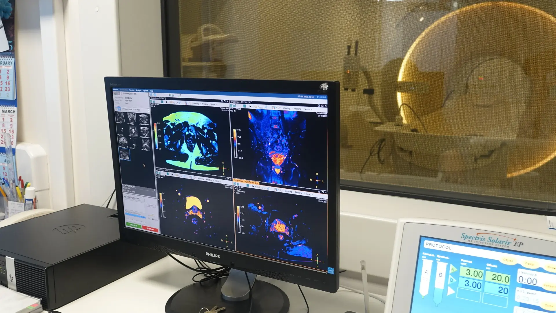
Decoding MRI images of the pelvis
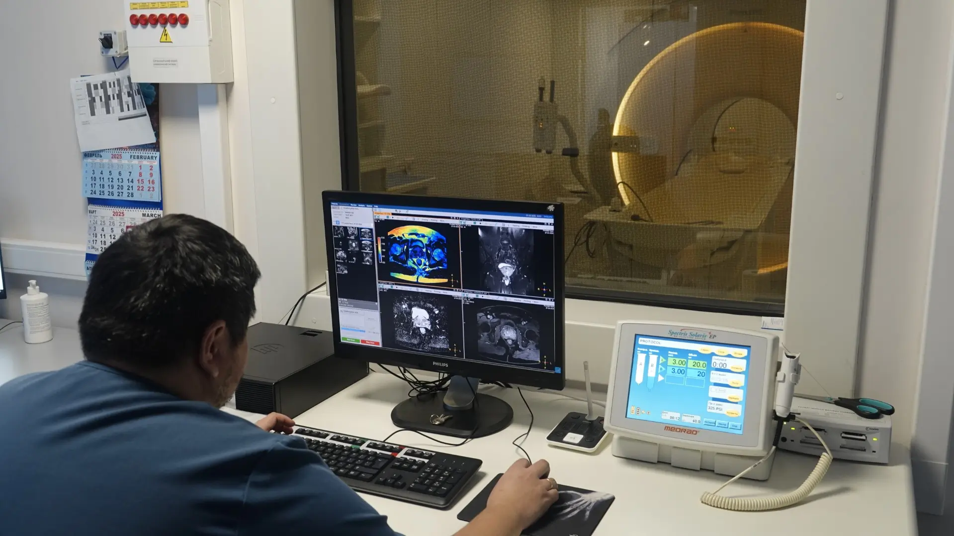
After conducting MRI of the female pelvic organs, the resulting images are analyzed by a radiologist. He carefully studies all the layered images, assessing the shape, size and structure of the organs, identifying even minor deviations from the norm. Particular attention is paid to such details as the thickness and homogeneity of the endometrium, the structure and location of the ovaries, the presence or absence of cysts, nodes, tumors, inflammatory or adhesive processes.
The doctor also assesses the condition of the surrounding soft tissues, vascular network, lymph nodes and adjacent organs, such as the bladder and rectum. In the case of using a contrast agent, the specialist additionally analyzes the nature of the blood supply to the tissues and the structure of the formations, which is especially important in differentiating benign and malignant processes.
Based on everything seen, the doctor makes a detailed conclusion in which he describes the detected changes and indicates which data require additional observation or consultation with a specialist. The results obtained are transferred to the patient and the attending physician to establish a final diagnosis and choose further treatment tactics.
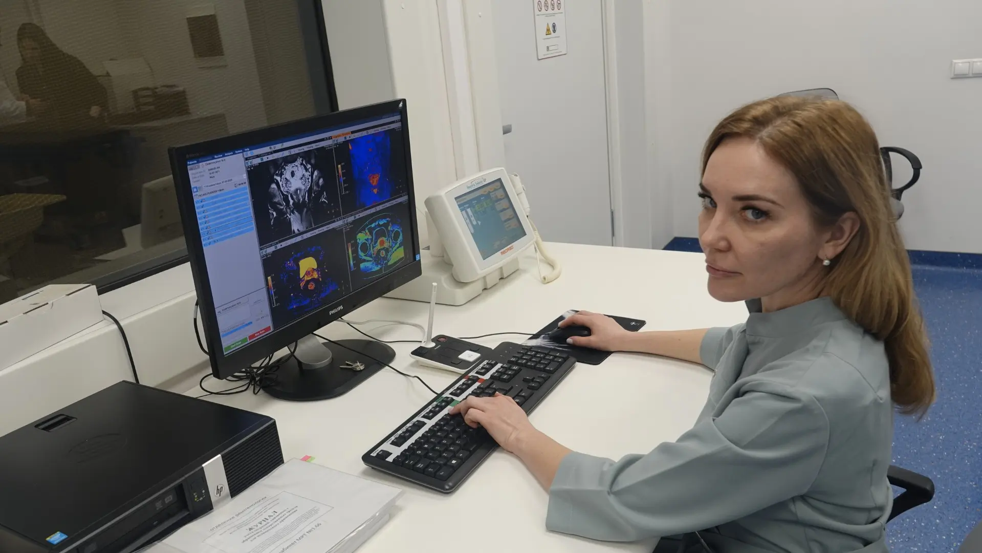
Safe diagnostic method
If you have gynecological problems and require an MRI of the pelvis, the N. Lopatkin Scientific Research Institute of Urology and Interventional Radiology is one of the best places to undergo such diagnostics. The institute uses modern visualization methods, with the possibility of an individual approach to each patient and the use of advanced equipment. Numerous positive reviews from patients emphasize the high qualifications of the staff, attentive attitude and clear organization of examinations, including consultations with specialized specialists. The institute is focused on a comprehensive approach to the diagnosis and treatment of gynecological diseases, and specialists have extensive experience in the field of women’s health.
MRI of the female pelvic organs is a modern, safe and highly informative diagnostic method that plays an important role in identifying and clarifying a wide variety of gynecological diseases. Due to the high image accuracy, MRI of the pelvis allows detecting even the smallest changes in tissues, identifying tumors, cysts, inflammatory and adhesive processes, as well as monitoring the effectiveness of treatment and recovery after surgery.
One of the main advantages of MRI of the pelvis is the absence of radiation exposure, which makes the study as gentle as possible for the female body. This is especially important during regular monitoring, preparation for pregnancy or IVF.
If you experience pain, menstrual irregularities or other alarming symptoms, do not delay contacting a doctor. If necessary, a specialist will recommend undergoing MRI of the pelvis to get a complete picture of the body’s condition and choose the most accurate and safe treatment strategy.
Before the examination, it is important to clarify with your doctor how to prepare for MRI of the pelvis. In case of contraindications, exclude the study. It is important to remember that preparation for MRI of the pelvis is an important stage that affects the accuracy of diagnosis and the effectiveness of the study.